
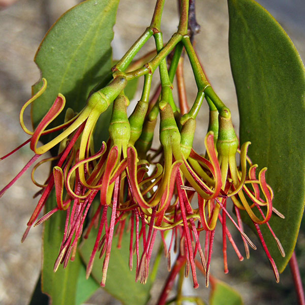
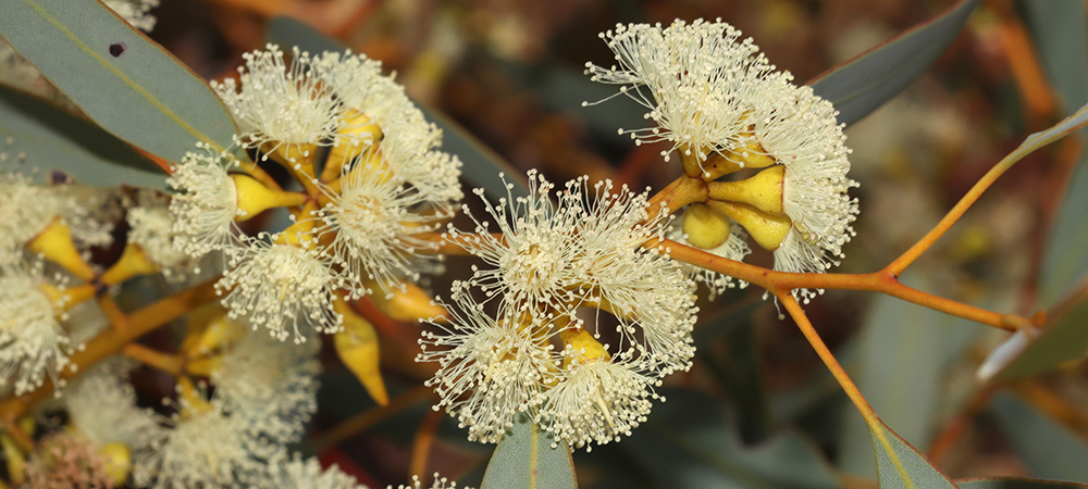
Ventilago viminalis, widespread in northern Australia. Photo: M. Fagg (APII).
For over 20 years, State Herbarium of South Australia botanist Jürgen Kellermann and colleagues Kevin Thiele (Canberra), Frank Udovicic and Neville Walsh (both Melbourne), are undertaking research on Rhamnaceae and revise the plant family for the Flora of Australia. Collaborators on the current research grant supporting this research by the Australian Biological Resources Study, Canberra, are Michelle Waycott, Ed Biffin, Korjent van Dijk from Adelaide and Catherine Clowes from Melbourne, as well as Francis Nge, who now works in Montpellier (France).

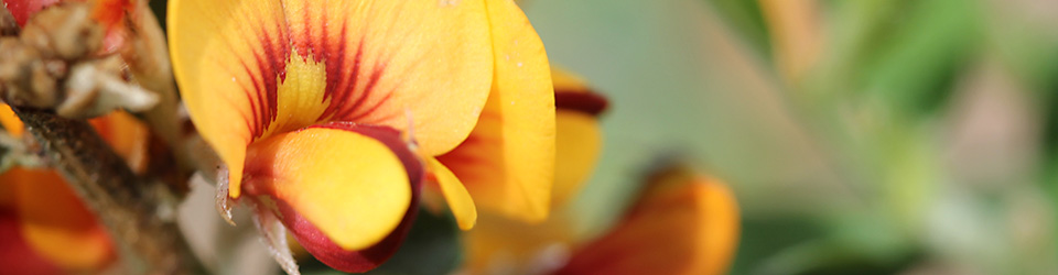
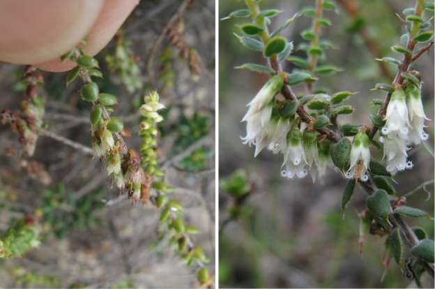
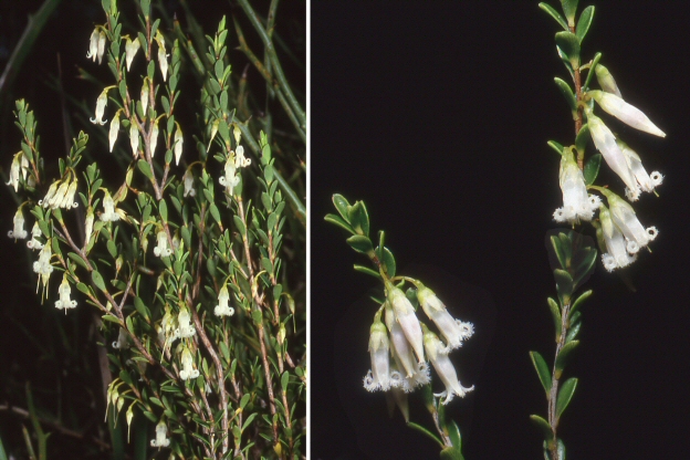
Polianthion wichurae from Western Australia. Photo: K.R. Thiele (APII).
Ramnaceae is a medium sized family of over 900 species and includes tropical and temperate trees, shrubs and some vines. It is cosmopolitan and species grow on all continents, except Antarctica. In Australia there are currently about 250 species recognised in 24 genera. There is a high level of endemism with at least 90% of Australian species occurring only on the continent. A significant number of species (approx. 30%) are classified as nationally rare or threatened.
This week, the treatments of 19 smaller genera (with 37 species) were published in the new online Flora of Australia. Most of these are widespread tropical and subtropical genera with only a few representatives in Australia: Alphitonia (5 species), Colubrina (2 spp.), Emmenosperma (2 spp.), Gouania (2 spp.), Hovenia (1 sp.), Noltea (1 sp.), Rhamnella (1 sp.), Rhamnus (2 spp.), Sageretia (1 sp.), Schistocarpaea (1 sp.), Ventilago (3 spp.) and Ziziphus (4 spp.). Some other genera are endemic to Western Australia or centred in this state: Blackallia (1 sp.), Granitites (1 sp.), Papistylus (2 spp.), Polianthion (4 spp.), Serichonus (1 sp.) and Siegfriedia (1 sp.). Finally, Discaria (2 spp.) occurs in eastern Australia and Tasmania, with related species in New Zealand and South America.
Work on the remaining five, larger genera (Cryptandra, Pomaderris, Stenanthemum, Spyridium and Trymalium) is in progress.

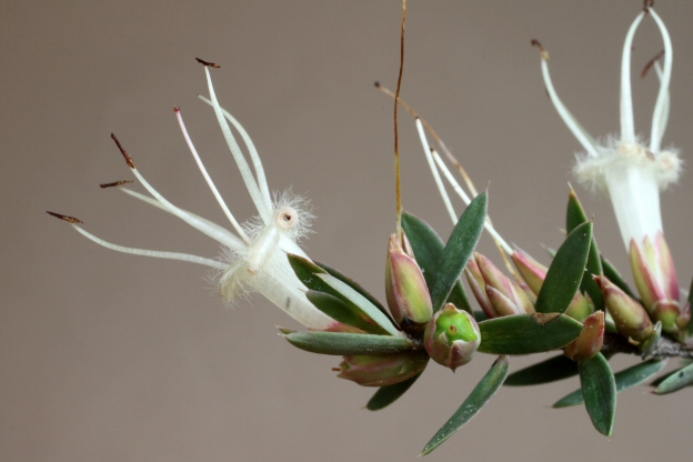
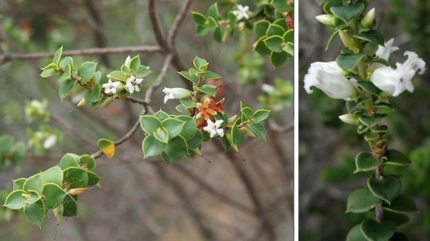
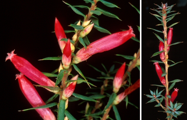
Granitites intangendus, a monotypic genus from Western Australia. Photo: K.R. Thiele (APII).
Written by State Herbarium botanist Jürgen Kellermann.


You must be logged in to post a comment.